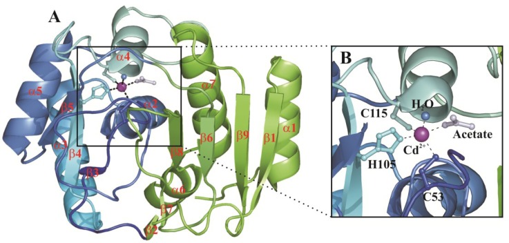
Figure 2.
(A) CDCA1-R1 overall fold. β-strands and α-helices are shown in cartoon representation and named as reported by [3]. Cd2+ is also depicted as purple sphere. The CDCA1 two-lobe architecture is highlighted by different colors: lobe 1 in blue and lobe 2 in green; (B) Enlarged view of the CDCA1-R1 active site. Cd2+ coordination sphere is shown.

