Figure 6.
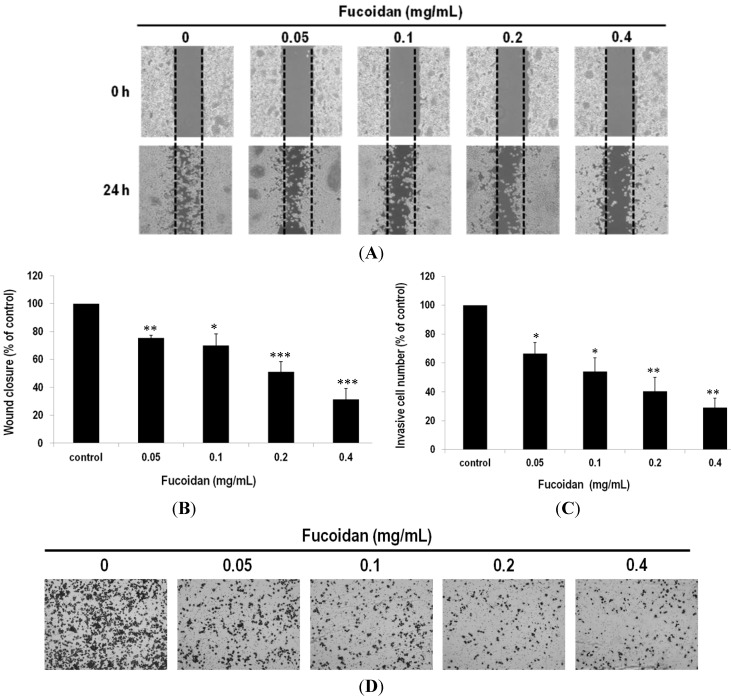
Suppression of migration and invasion of lung adenocarcinoma cells by fucoidan. (A) Representative photographs of three independent experiments, showing a dose-dependent inhibition of migration after treatment of fucoidan (24 h). Images of wound closures (10× magnification); (B) Black dotted lines indicate the wound edge. The cell-free areas invaded by cells (across the black dotted lines) were quantified by three random fields as shown in the lower panels; (C) The invasiveness of the LLC cells were quantified by counting the stained cells that invade into the porous polycarbonate membrane; (D) Invasiveness of the LLC cells treated with fucoidan. The LLC cells were pretreated with fucoidan for 24 h and then seeded onto the transwell chamber. Photographs were taken by an inverted microscope with 10× magnification. Data were derived from three independent experiments and presented as mean ± SEM. * p < 0.05, ** p < 0.001, *** p < 0.0001 when compared to the vehicle-treated cells.

