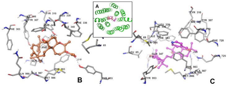
Figure 5.
XP-Glide predicted binding mode of SSJ26 and SSJ32 with homology modeled P-gp. (A) Docked poses of SSJ26 and SSJ32 into drug binding sites of human P-gp. Backbone of human P-gp is depicted as gray ribbons viewed from the intracellular side of the protein looking into the internal chamber. SSJ26 and SSJ32 are represented as tube models with the same color scheme below; (B) Binding mode of SSJ26 within human P-gp. Important residues are depicted as tubes with the atoms colored as carbon—gray, hydrogen—white, nitrogen—blue, oxygen—red, sulfur—yellow, whereas SSJ26 is shown as ball and stick model with the same color scheme as above except carbon atoms are represented in orange. Dotted black lines indicate hydrogen bonds; (C) Binding mode of SSJ32 within the human P-gp. The color scheme is same except carbon atoms are represented in purple.

