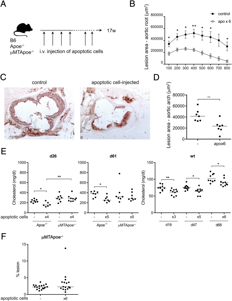
Fig. 4.
Apoptotic cell injections protect against lesion development and lower cholesterol in a B-cell-dependent way. Apoe−/− and C57BL/6 (WT) mice were injected six times with apoptotic cells during disease progression, as indicated by the scheme shown in A. Aortic lesions in the aortic root, assessed by oil red O staining, showed reduced lesions in apoptotic cell-injected Apoe−/− compared with uninjected controls (C), quantified as lesion area (B). Lesion in aortas assessed by Sudan IV staining in D and F. Median and individual mice [n = 6–7 (D); n = 16–17 (F)] are plotted. Cholesterol levels, measured using enzymatic colorimetric assays, in apoptotic cell-injected (×3, ×4, ×5, or ×6) Apoe−/−, μMTApoe−/−, or WT mice compared with uninjected (−) age-matched controls at indicated days after the first injection (E). Median (Left and Middle) or mean (Right) and individual mice (n = 6–10) are plotted. Data shown from one experiment in which n = 5–8 (B and C), a separate experiment in which n = 7 (D), representative of 2 experiments in which n = 6–10 (E), pooled from two and representative of three experiments in which n = 3–9 (μMTApoe−/− in E) or pooled from three experiments (F). *P < 0.05 and **P < 0.01 by a Mann–Whitney U test or Student’s t test in E (Right).

