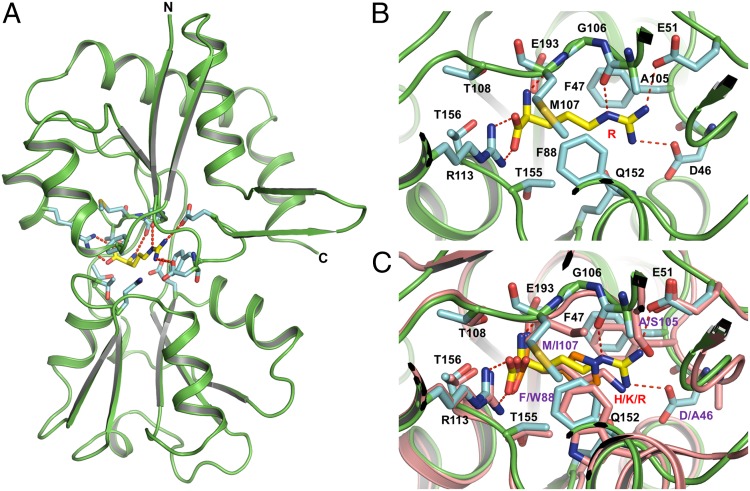
Fig. 2.
Overview structure of the substrate-binding protein ArtI complexed with Arg. (A) ArtI is colored in green. The residues represented in cyan sticks interact with Arg mostly through hydrogen bonds and van der Waals force. Arg is shown in ball-and-stick models. Hydrogen bonds are represented as red dashed lines. (B) Stereoview of a detailed ribbon diagram of the Arg binding site of ArtI. Representative models of the residues from the substrate binding site as well as Arg are the same as in A; hydrogen bonds are represented as red dashed lines. (C) Stereoview of the structural alignment between ArtI and ArtJ of the ArtJ-(MP)2 transporter from G. stearothermophilus (PDB ID codes 2Q2A, 2PVU, and 2Q2C). ArtI and ArtJ are colored in green and pink, respectively. Lys, Arg, and His are shown in ball-and-stick models. In ArtJ, residues that mediate substrate binding are shown in pink sticks.

