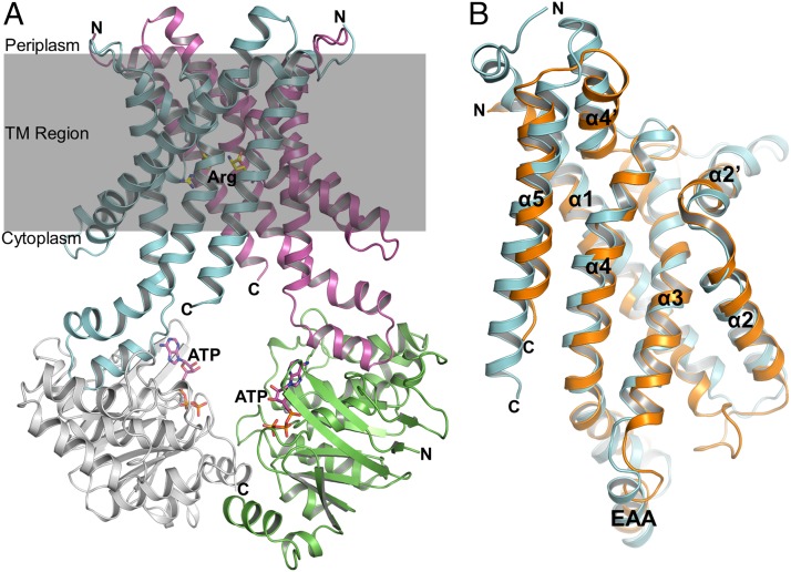
Fig. 3.
Overview structure of the Art(QN)2 complex in an inward-facing conformation. (A) Two transmembrane subunits (ArtQ) are colored in cyan and magenta, and two nucleotide-binding subunits (ArtN) are colored in gray and green. Arg and ATP molecules are shown in ball-and-stick models. (B) Stereo views of the structural alignment between ArtQ and MetI of the methionine uptake system Met(NI)2 of Escherichia coli (PDB ID code 3DHW). MetI and ArtQ are colored in orange and cyan, respectively.

