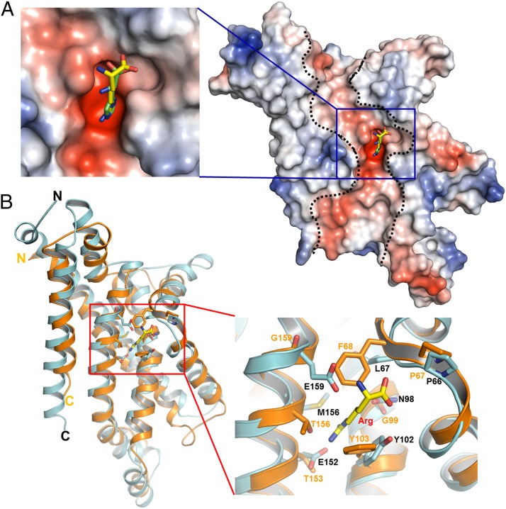
Fig. 5.
A highly negatively charged substrate binding pocket lies in the middle of the protein. (A) ArtQ contains a highly negatively charged electrostatic potential tunnel (indicated by dashed lines) reaching from the periplasmic side to the cytoplasmic side. A highly negatively charged substrate-binding pocket, indicated by a blue square, lies in the middle of the protein. Arg is shown in ball-and-stick representation. (B) Stereo views of the structural alignment of the substrate-binding site of ArtQ with the corresponding region of MetI of the methionine uptake system Met(NI)2 from E. coli (PDB ID code 3DHW). MetI and ArtQ dimers are colored in orange and cyan, respectively. The residues interacting with substrate as well as Arg are shown in ball-and-stick models.

