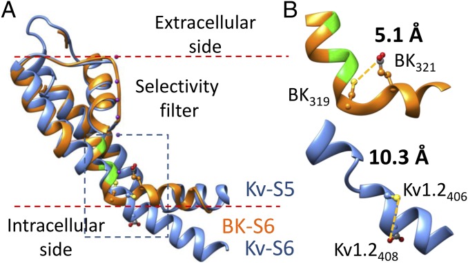
Fig. 5.
A hypothetic structure of the BK PGD based on the Kv1.2 crystal structure and functional results from our previous and current studies. (A) The hypothetic structure of BK PGD (orange) is superimposed with the Kv1.2 structure (blue). Three pore-lining residues of BK channel (A313, A316, and S317) are colored in green. The S5 of the BK structure is omitted for clarity. K+ ions are rendered as purple balls. Lipid membrane is delimited by red dotted lines. The boxed region is magnified in B to show the structure around the YVP/PVP motif. Side chains of BK319, BK321, Kv1.2406, and Kv1.2408 (equivalent to Shaker474 and Shaker476) are rendered as ball and chain. Notice that Kv1.2406 and Kv1.2408 are not on the same surface of the Kv S6 helix.

