Fig. 3.
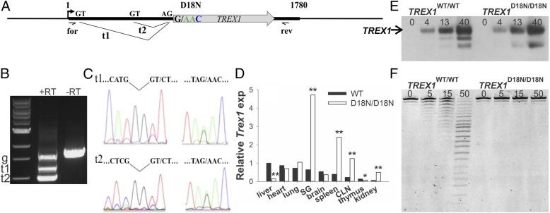
The TREX1 D18N allele is expressed, processed, and translated. (A) The TREX1 single exon gene is shown with the two donor (GT) and acceptor (AG) sequences. Transcripts 1 (t1) and 2 (t2) are generated from the two donor sites. (B), TREX1 cDNA products were generated from TREX1WT/D18N mouse liver RNA using forward and reverse primers positioned as shown in A. Reactions containing reverse transcriptase (RT)-generated t1 and t2. The genomic (g) TREX1 sequence was generated in reactions ±RT. (C) Sequencing of t1, t2, and g bands in B demonstrates appropriate t1 and t2 splicing (Left) and expression of TREX1 D18N and WT alleles (Right). (D) TREX1 expression in mouse tissues (six animals of each genotype). SG, salivary gland; CLN, cervical lymph node. (E) Western blot of TREX1 protein (ng) purified from mouse tissues. The position of migration of TREX1 is indicated. The band identified as TREX1 D18N was excised from a gel and confirmed by mass spectroscopic analysis (F). Activity assay of TREX1 protein (pg) purified from mouse tissues. *P < 0.05; **P < 0.001.

