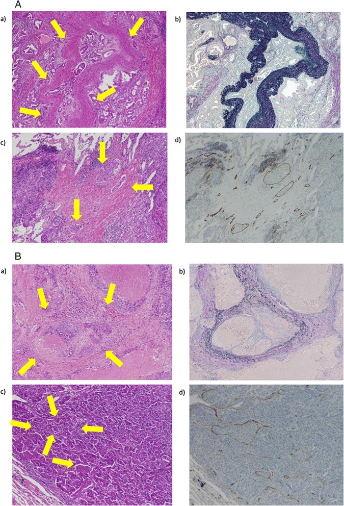
Figure 1.

Detection of vascular invasion by hematoxylin-eosin (HE) staining and other staining methods. A: Representative case of vascular invasion by HE staining and confirmed using other staining methods. a) HE staining for the diagnosis of blood vessel invasion (v). b) Elastica van Gieson staining to confirm v in the same patient. c) HE staining identified multiple instances of lymphatic vessel invasion (ly). d) D2-40 staining to confirm ly in the same patient. B: Representative case of false-negative vascular invasion identified on HE staining. a) v could not be determined on HE staining. b) Elastica van Gieson staining showed v in the same patient. c) Lymphatic vessel invasion (ly) could not be determined on HE staining. d) D2-40 staining showed multiple instances of ly in the same patient.
