Figure 3.

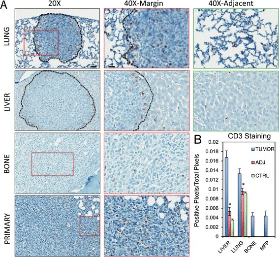
T lymphocytes are recruited to liver and lung metastases from breast cancer. Paraffin-embedded sections from primary breast tumors, bone specimens, lung specimens and liver specimens obtained following experimental metastasis assays were subjected to immunohistochemical staining with anti-CD3 antibodies. (A) Representative images from 20X, 40X magnifications for each site are shown. 40X images were taken at the margin of the lesions (40X margin) and in more distal regions (40X adj.). Scale bar represents 40 μm (20X) or 20 μm (40X) and applies to all panels of the same magnification. (B) Positivity of CD3 staining (expressed as a ratio of positive pixels over the total pixels per field) quantified inside lesions (TUMOR), in the adjacent tissue (ADJ) or in control (CTRL) samples without any lesions. Lymphocyte T expression and recruitment is mostly associated with lung or liver metastasis (*: P <0.001).
