
Figure 6.
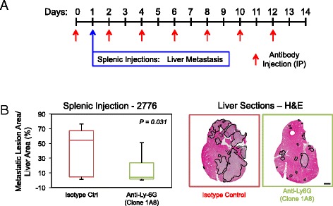
Ly-6G + cell depletion decreases the establishment and growth of liver metastases. (A) Schematic depicting the experimental protocol for depletion of Ly-6G+ cells. (B) Paraffin-embedded liver sections were obtained following completion of the experimental metastasis assays and subjected to immunohistochemical staining with anti-Ly-6G antibodies. Statistically significant decreases in Ly-6G+ cells that infiltrate the liver were observed when the neutrophil-depleted cohort was compared with the isotype control cohort (P = 0.045). Representative images of Ly-6G-stained liver sections from each cohort. (C) Quantification of the tumor burden (tumor area/tissue area) within the cardiac liver lobe following splenic injection of 2776 liver-aggressive cells. Statistically significant decreases in liver-metastatic burden were observed when the isotype control cohort was compared with the neutrophil-depleted cohort (P = 0.031). Representative images of hematoxylin and eosin (H&E)-stained liver sections exhibiting the liver-metastatic burden in each cohort. Dotted lines circumscribe breast cancer metastatic lesions within the liver. Scale bar represents 2 mm and applies to all panels. IP: intraperitoneal injection.