
Figure 8.
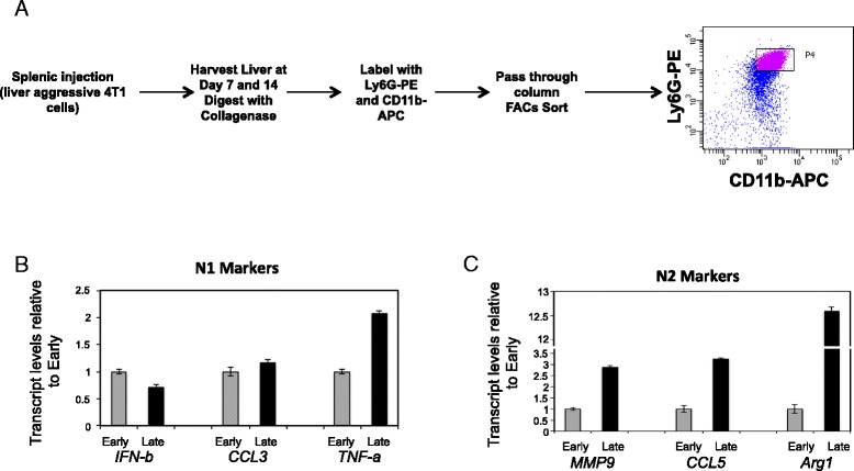
N2-polarized neutrophils are recruited to the invasive front of liver metastases over time. (A) Schematic depicting the experimental protocol to isolate neutrophils from metastasis-bearing livers. (B) Quantitative real-time PCR analysis was performed for IFN-β, CCL3 and TNF-α as markers for anti-tumorigenic (N1)-polarized neutrophils, normalized to total Gapdh levels, in neutrophils isolated from early (7 days) or late (14 days) time points post splenic injection with the 2776 liver-aggressive cell line. The data is depicted as fold expression relative to early time point and is representative of four independent experiments performed in triplicate. (C) Quantitative real-time PCR analysis was performed for MMP9, CCL5 and Arginase 1 as markers for pro-tumorigenic (N2)-polarized neutrophils, normalized to total Gapdh levels, in neutrophils isolated from early or late time points post splenic injection with the 2776 liver-aggressive cell line. The data is depicted as fold expression relative to early time point and is representative of four independent experiments performed in triplicate.