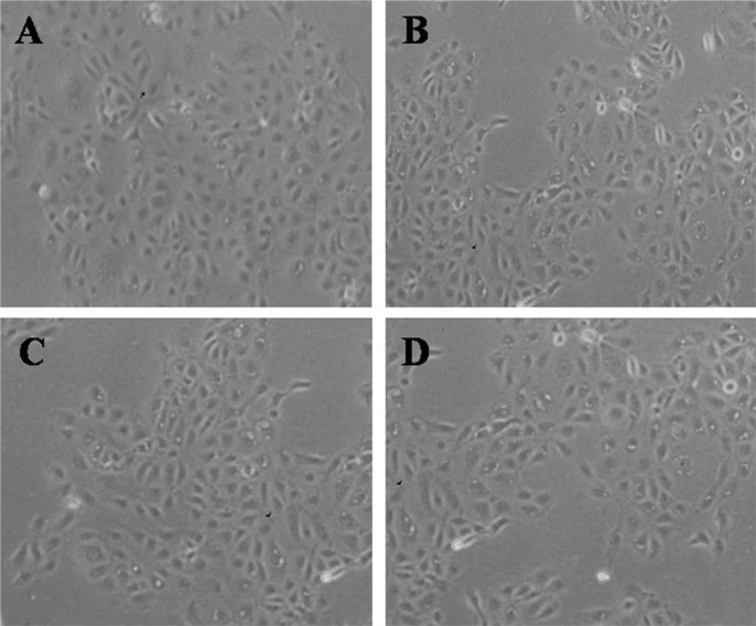
Figure 5. Inverted phase-contrast micrographs of HCECs after exposure to 0.5% CMC, 0.3% HA and 0.1% HA (original magnification, ×200).
Many epithelial cells are visible in control culture media (A). The number of HCECs were decreased and more detached from the culture media, in the case of exposure to 0.5% CMC (B), 0.3% HA (C) and 0.1% HA (D) compared with control.

