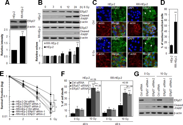
Figure 1. Depletion of ERp57 sensitizes radioresistant HEp-2 cells.

(A) Lysates of control HEp-2 and RR-HEp-2 cells were immunoblotted with an anti-ERp57 antibody. (B) Control HEp-2 and RR-HEp-2 cells were treated with 6 Gy radiation and cultured for the indicated periods. Expression levels of ERp57 were quantified using ImageJ software from 3 independent experiments (lower panel, A and B). **P < 0.01 compared with HEp-2 (A) or untreated HEp-2 cells (B) and × denotes no significance compared with untreated RR-HEp-2 cells (B). (C and D) HEp-2 or RR-HEp-2 cells untreated (Ctrl) or treated with 6 Gy radiation for 24 h were stained with anti-ERp57 antibody (green), Alexa 568 phalloidin (red), and DAPI (blue). The asterisks and arrows indicate fragmented nuclei and ERp57 nuclear localization, respectively (C). Nuclear-localized ERp57 in HEp-2 and RR-HEp-2 cells was quantified using CellProfiler software (≥100 cells for each dataset). n = 3; **P < 0.01 compared with HEp-2 cells (D). (E–G) RR-HEp-2 cells were transfected with 100 nM of control siRNA, ERp57 siRNA-1, or ERp57 siRNA-2, respectively. After 48 h, the cells were treated with each dose of radiation, as indicated, (E) or 10 Gy radiation (F and G). # indicates the ratio for % of cell death of irradiated siRNA control cells / % of cell death of irradiated ERp57-depleted cells. The clonogenic survival fraction was determined by clonogenic assay (E). Cell viability was determined via FACScan flow cytometry, and data are presented as the percentage of PI-positive cells (F). Cells were analyzed by immunoblotting with anti-cleaved PARP and anti-ERp57 antibodies (G). β-actin was used as loading control. The data represent typical results and are presented as mean ± standard deviation of 3 independent experiments; *P < 0.05 or **P < 0.01 compared with irradiated siRNA control cells, respectively (E and F).
