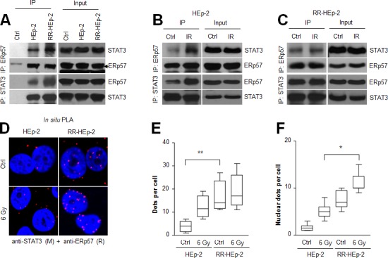
Figure 2. The physical interaction between ERp57 and STAT3 is increased in radioresistant HEp-2 cells.

(A-C) Indicated cell lysates were immunoprecipitated with anti-ERp57 antibody (upper panel), anti-STAT3 antibody (lower panel) or their respective control immunoglobulin G (IgG: A, Ctrl lane) antibody and immunoblotted with anti-STAT3 or anti-ERp57 antibody. The combined HEp-2 and RR-HEp-2 cell lysates were used as the experimental control (A, Ctrl lane). (B–E) HEp-2 cells or RR-HEp-2 cells were untreated (Ctrl) or treated with 6 Gy radiation (IR) for 12 h. (D-F) The cells were fixed and incubated with mouse anti-STAT3 together with rabbit anti-ERp57, followed by in situ PLA analysis. Representative confocal images of cells with PLA-positive signals are shown (D). Dots per cell were counted using CellProfiler (E and F). The data represent typical results and are presented as mean ± standard deviation from 3 independent experiments; **P < 0.01 compared with untreated HEp-2 cells (E) and *P < 0.05 compared with irradiated HEp-2 cells (F).
