
Figure 2. Silvestrol leads to depletion of non-irradiated LCL in co-cultures while permitting expansion of T and NK cells.
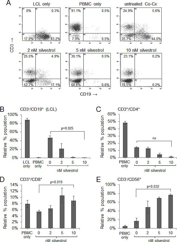
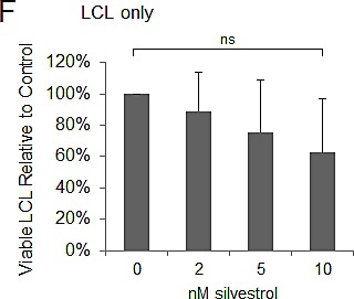
CoCx (N=3) were created by mixing non-irradiated LCL with equal numbers of autologous peripheral blood mononuclear cells (PBMC). CoCx or PBMC alone were incubated in the presence of 10 U/ml IL-2 and given a single dose of 0 (vehicle only), 2, 5, or 10 nM silvestrol. Flow cytometric analysis was conducted on day 10. For all results, live events were gathered by gating on cells negative for the LIVE/DEAD stain. (A) Representative flow cytometry dot plots of mononuclear cells from CoCx. Cells were stained for CD3 (y-axis) and CD19 (x-axis) and gated on live events. LCL (CD3−/CD19+) are shown in the bottom right quadrant of each panel. (B-E) Data are expressed as percentage of total viable population expressing: (B) CD3−/CD19+ (LCL); (C) CD3+/CD4+ (helper T-cells); (D) CD3+/CD8+ (cytotoxic T cells); (E) CD3−/CD56+ (NK cells). All results are averages of three individual CoCx; ns = not significant. (F) Four different LCL were cultured using the same conditions as in the above co-cultures, but without the addition of PBMCs, and incubated with a single dose of silvestrol. Viable LCL were enumerated by flow cytometry, using cell counting beads and gating on cells negative for the LIVE/DEAD stain, and are shown relative to the untreated (vehicle) control. Differences with silvestrol treatment (0 versus 10 nM) were not significant (ns).