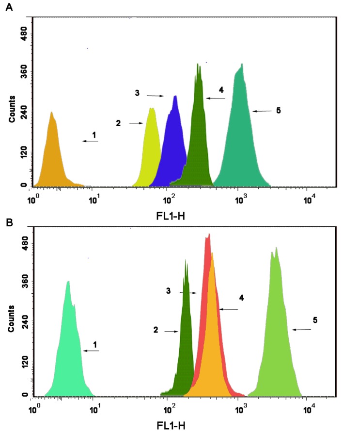
Figure 5. Drug content of the mitochondrial fraction in MCF-7 cells.
(A) and MCF-7/Adr cells (B) after applying different formulations, as determined by flow cytometry. The abscissa indicates the fluorescence intensity of a 6-coumarin formulation internalized by the cancer cells, and the ordinate represents the cell counts. Notes: A1 and B1, blank control; A2 and B2, free 6-coumarin; A3 and B3, 6-coumarin nanoparticles; A4 and B4, targeted 6-coumarin nanoparticles; A5 and B5, functional 6-coumarin nanoparticles.

