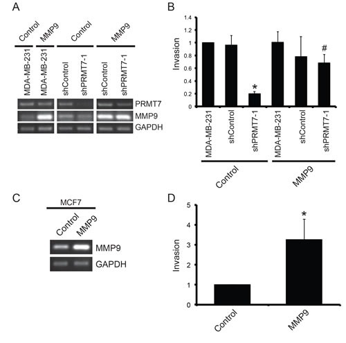
Figure 7. Overexpression of MMP9 rescues the loss of invasion resulting from PRMT7 depletion.

MDA-MB-231 cells stably depleted of PRMT7 using shRNA were transiently transfected with an empty vector (Control) or a vector containing MMP9 cDNA. Parental MDA-MB-231 (MDA-MB-231) and cells expressing a non-targeting shRNA cells were used as controls. Total RNA was collected 24 h post transfection and used to generate cDNA to assess PRMT7 and MMP9 expression levels (A). GAPDH served as a loading control. To examine the effect of MMP9 overexpression on cell invasion, MDA-MB-231 cells (MDA-MB-231), cells stably expressing a control shRNA (shControl) and MDA-MB-231 cells stably depleted of PRMT7 were transfected for 24 h as described above. Twenty-four hours post transfection, cells were re-plated at equal numbers into Transwell chambers containing a Matrigel layer and incubated for an additional 24 h. Cells that passed through the chambers were counted (B). Data represents the mean ± standard error of three independent experiments (*p < 0.05 compared to MDA-MB-231 cells stably expressing a control shRNA, #p < 0.05 compared to MDA-MB-231 cells stably depleted of PRMT7 transfected with an empty vector). MCF7 cells were transiently transfected with an empty vector (Control) or a vector containing MMP9 cDNA. Total RNA was collected 24 h post transfection to assess expression. PCR analysis of cDNA generated from total RNA using MMP9 primers shows expression levels (C). GAPDH serves as a loading control. To examine the effect of MMP9 overexpression on cell invasion, MCF7 cells were transfected for 24 h as described above and then replated at equal numbers into Transwell chambers containing a Matrigel layer and incubated for an additional 72 h. Cells that passed through the chambers were counted (D). Data represents the mean ± standard error of five independent experiments (*p < 0.05).
