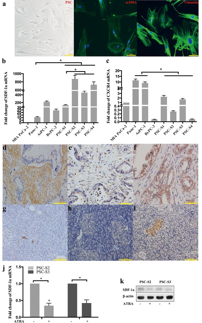
Figure 1. SDF-1α and CXCR4 expression in PSCs and PCCs.
Activated primary PSCs isolated from pancreatic cancer tissues were verified by immunofluorescence staining for α-SMA and vimentin (a); SDF-1a mRNA (b) and CXCR4 (c) mRNA expression was examined by RT-qPCR. The expression of the target mRNA was normalized to that of β-actin mRNA. The values are expressed relative to 1.00 for expression in MIA PaCa-2 cells. The bars represent the mean of three independent experiments ± SE. *, P<0.05. In the four resected pancreatic cancer samples used for PSC isolation, α-SMA, SDF-1α and CXCR4 were detected by immunohistochemistry in pancreatic cancer tissues (d, e and f) and distant normal pancreatic tissues (g, h and i). Two primary PSCs were harvested and analyzed for SDF-1α expression by RT-qPCR (j) and Western blot (k).

