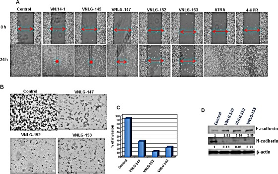
Figure 5. Anti-migratory and anti-invasive potential of NRs.

(A) Effect of NRs treatment (5 μmol/L) on PC-3 cell migration was assessed by wound healing assay after 24 h of wound formation. Representative photomicrographs of initial and final wounds are shown at 100x magnification. (B) Effect of NRs on PC-3 cell invasion was evaluated by transwell migration assay. PC-3 cells were seeded on matrigel coated boyden chamber and treated with NRs (5 μmol/L, 24 h). Photographs represent the extent of cell invasion in each of the treated cells. (C) Western blot analysis of the effect of NRs on the expression of cadherins in PC-3 cells. Cells were treated with indicated compound 20 μmol/L for 24 h. Total cell lysates were separated by SDS-PAGE and probed with E- and N-cadherin antibodies. Vehicle treated cells were included as a control and all blots were reprobed for β-actin for equal protein loading and transfer.
