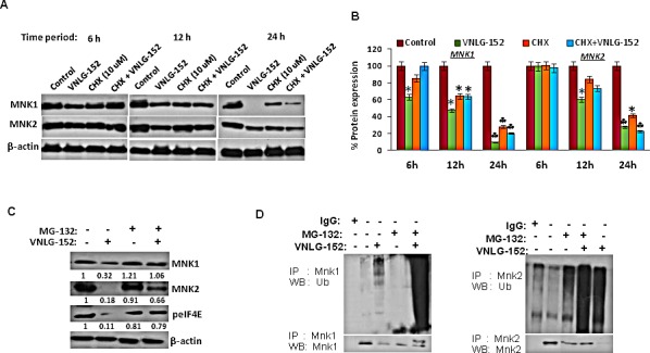
Figure 8. VNLG-152 induced MNK1/2 degradation by ubiquitin proteasomal pathway in LNCaP cells.

(A) Western blot analysis of the expression of MNK1/2 in LNCaP cells treated with VNLG-152 (20 μmol/L) in the presence or absence of CHX (10 μmol/L) at 6, 12 and 24 h of treatment. (B) Densitometric analysis of the expression of MNK1/2 in different groups *, P< 0.05; ♣, P<0.01 compared with vehicle treated control. (C) Western blot analysis of the expression of MNK1/2 and peIF4E in LNCaP cells treated with VNLG-152 (20 μmol/L) in the presence or absence of MG-132 (5 μmol/L). Vehicle treated cells were included as control and all blots were reprobed for β-actin to ensure equal protein loading. (D) LNCaP cells were treated with 20 μmol/L of VNLG-152, 5 μmol/L of MG-132, and combinations for 24 h. MNK1/2 protein was immunoprecipitated with MNK1/2 antibody (mouse) respectively and the precipitated protein was subjected to western blot analysis with anti-ubiquitin antibody (Ub) (upper panel). The same blot was used to detect MNK1/2 protein with anti-MNK1/2 (rabbit) antibody after stripping (lower panel).
