Figure 5.
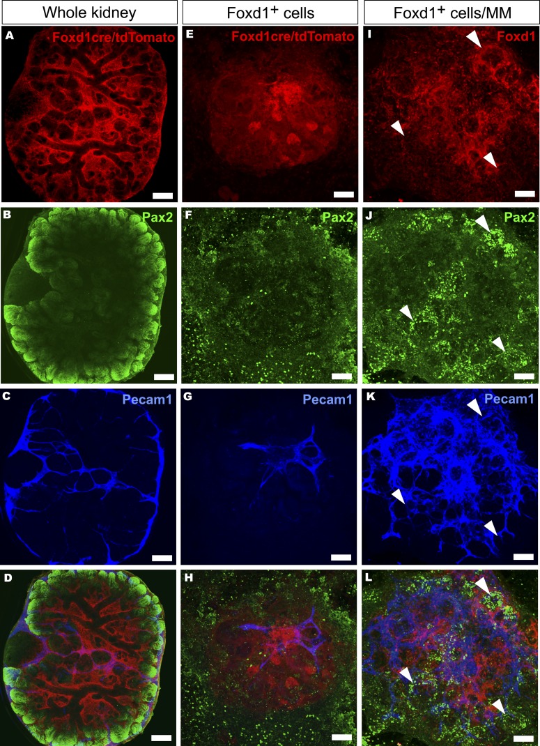
Interaction of Foxd1+-derived stromal cells with embryonic kidney mesenchymal progenitor cells is critical for endothelial network and nephron development. (A–D) The fate of the Foxd1cre+/floxed Rosa26tdTomato-marked stromal progenitor cells (in red), the kidney tubular cells (in green, Pax2+), and endothelial cells (in blue, Pecam1+) is mapped in whole-kidney time-lapse organ culture in the embryonic kidney mesenchyme (MM). (E–L) Alternatively, the stromal cell behavior is addressed in FACS-purified, homotypic Foxd1+ cells aggregates (E–H) or when the extracted Foxd1+ cells were recombined with the rest of the embryonic kidney MM cells (I–L). The Foxd1cre-marked tdTomato+ cells (in red) are found around the nonstained ureteric tree (A and D) and the developing nephrons are depicted with Pax2 staining (B and D, in green), resembling the situation in vivo. The endothelial cells establish a network within and around the kidney as highlighted by Pecam1 expression (C, in blue). (E–H) The FACS-purified dMM-derived Foxd1cre-marked tdTomato+ cells are aggregated for homotypic cultures and subjected to the eSC tubule inducer. In such a setting, the FoxD1+ cell aggregates survive (E) but no tubules appear, as illustrated by the failure in epithelial tubular organization and weak Pax2 expression (F, in green). Endothelial Pecam1 expression (G, in blue) is also limited in these cells. (J and K) The presence of the Foxd1+ cells (in red) with the rest of the MM cells promoted both Pax2 expression (J, in green; arrowheads) and differentiation of the Pecam1+ endothelial network (K, in blue). Culture time is 5 days. Bar, 200 µm.

