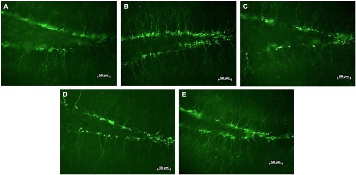
Figure 1.
DCX-positive cells in the subgranular zone of the hippocampal dentate gyrus of female mice. Photomicrographs of representative responses when females were exposed to a water control (A), male CD-1 urine (B), male BALB/c urine (C), male BALB/c urine mixed with 1 μg/μl r-darcin (D) or female CD-1 urine (E). Mean cell counts for each treatment are shown in Figure 2A.

