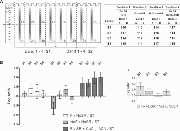
Figure 2.
(A) Left panel: Equal amounts of a HepG2 cell lysate are divided over 16 lanes of two gels, whereby a loaded lane is alternated with an empty one to allow easy cutting of the gel (schematic of one gel is shown). After electrophoresis, the gels are cut around the 50 kDa marker to create a high (A) and low (B) molecular weight fraction. The different lanes were excised and in-gel digestion was performed on the different gel bands according to different conditions and pooled for each replicate after labeling. Right panel: Condition 1—digestion of a fixed and SR-stained gel, marked as the standard (ST) protocol. Condition 2—digestion of a fixed, nonstained gel. Condition 3—digestion of a nonfixed, nonstained gel. Condition 4—ST supplemented with 1 mM CaCl2 and 5% ACN. Extracted peptides from bands 1 to 4 were labeled and pooled according to the presented schedule, together forming four high and four low molecular weight samples. S1–S4: different replicates. (B) Each bar represents the average and SD of all the reporters of one of the four replicas. The different conditions are compared to the standard situation where the gel is fixed and SR stained. Despite a large variation, ratios indicate that SR has a negative effect on peptide recovery and fixation a positive influence. Addition of CaCl2 and ACN during trypsin digestion increases the peptide yield over sevenfold in average. Asterisk: The positive effect of gel fixation is verified by three of four replicates from the “fixation no SR/no fixation no SR” ratios.

