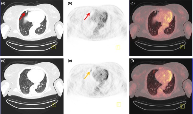
Figure 1.

PET/CT scan of a 60-year-old female with a tumour anterior in the right lung, easily visible on CT (a). However, on PET/CT, FDG uptake was hardly visible in the area of the tumour (b,c). Taking a closer look at the slices above the tumour (d–f), moderately focal increased FDG uptake was visible here (SUVmax = 4). This uptake corresponds to the tumour, but as a result of respiratory movements during PET acquisition, there is a misalignment between CT and FDG-PET. The patient was diagnosed with a carcinoid tumour, which explains the relatively low FDG uptake.
