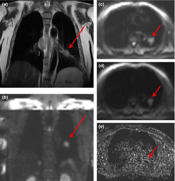
Figure 4.

MR scans of a 54-year-old patient with adenocarcinoma in the left lung. During initial examination on coronal T2-weighted images, (a) primary tumour (red arrow) is seen as being isointense to soft tissue. Correspondent coronal DW image displays tumour as a hyperintense zone (b). On the transaxial images, the SI (signal intensity) of the primary tumour (on b-value of 50 and 1000 s mm−2) is seen to stay relatively high and only decrease slightly together with lesion diameter if b-value is elevated (c and d). On ADC (e) map, the primary lesion shows low SI, corresponding to the malignant nature of the tumour.
