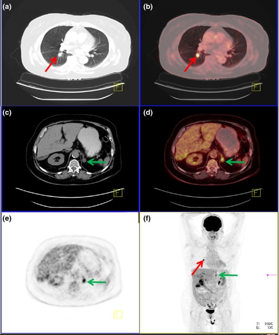
Figure 5.

PET/CT scan of a 66-year-old female with a central tumour in the right lung (red arrow), easily visible on CT (a). Tumour was highly FDG-avid indicative of malignancy (SUVmax = 16) (b). An enlarged left adrenal gland was visible on CT (green arrow) (c) and similar to the primary tumour highly FDG-avid, corresponding to a metastasis (SUVmax = 9) (d,e). Both findings can be seen on the multi-intensity projection (MIP, f) and was confirmed to be adenocarcinoma.
