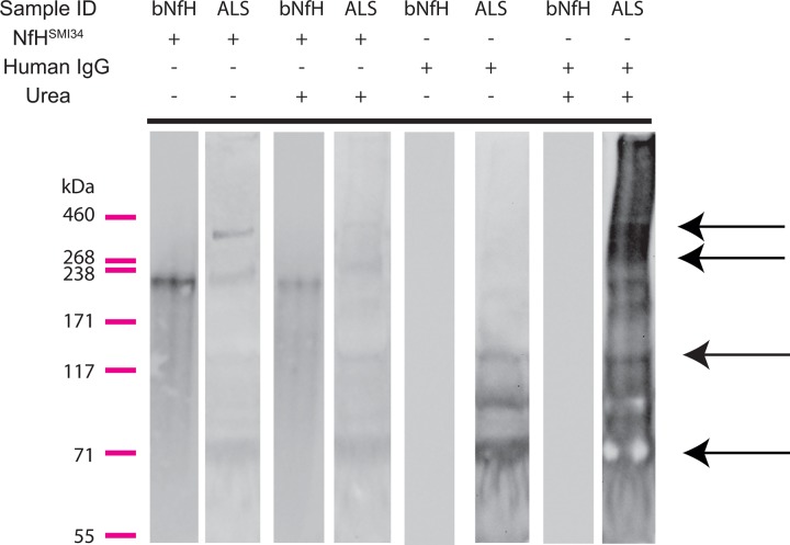
Figure 4.
Immunoblots of plasma samples from patients with amyotrophic lateral sclerosis (ALS) and of purified bovine neurofilament heavy chain protein (NfH) proteins. NfH bands represent high molecular weight (MW) aggregates (238–460 kDa), monomers and NfH fragments (bands below ∼205 kDa) in plasma samples from patients with ALS (the second lane from the left of the panel), while only a monomer band for purified bovine NfH protein is displayed bovine NfH (bNfH, the first lane from the left of the panel). Urea partially dissolved the high MW NfH from ALS plasma as shown previously in superoxide dismutase1 (SOD1)G93A mice (the fourth lane from the left side of the panel), but had no effect on bNfH (the third line form the left side of the panel; refer J Neurosci Methods). After stripping of the NfH antibodies, the blot was reprobed with antihuman IgG (the four lanes from the right side of the panel). In ALS samples, multiple bands showing intense staining for human IgG were present at the level of NfH high MW aggregates, NfH monomers and NfH endogenous fragments and (black arrows), but not in the bNfH lanes (the second and fourth lanes on the right side).

