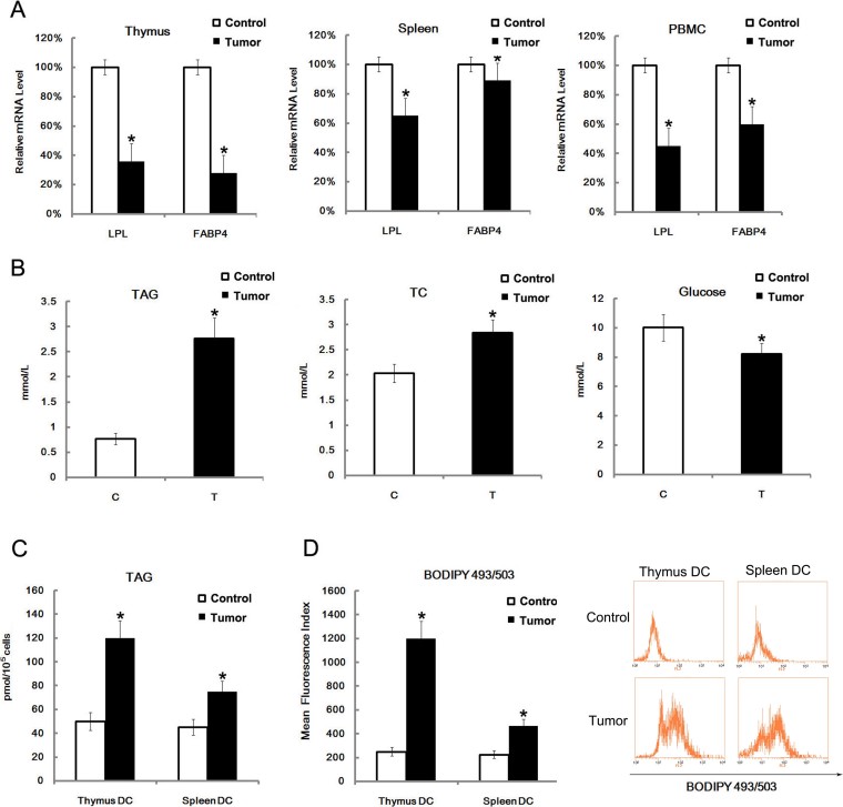
Figure 3. LPL and FABP4 levels were down-regulated, TAG was up-regulated and lipid content in DC was increased in radiation-induced thymic lymphomas.
Thymus, spleen and PBMC were isolated from mice with radiation-induced thymic lymphomas. Expression of LPL and FABP4 were assayed by qRT-PCR analysis of purified total mRNA (A). The TAG, TC and glucose levels in serum were assayed. Data are presented as the mean ± s.d. of three independent samples (B). The thymus and spleen were isolated, and thymic and splenic DCs were purified by sorting CD11c+ positive cells. The disrupted cells were then assayed for TAG level (C). Purified thymic and splenic DCs were stained with BODIPY493/503 (D). All data are presented as the mean ± s.d. of three independent experiments. *P < 0.05.

