. 2015 Mar 27;4:e06119. doi: 10.7554/eLife.06119

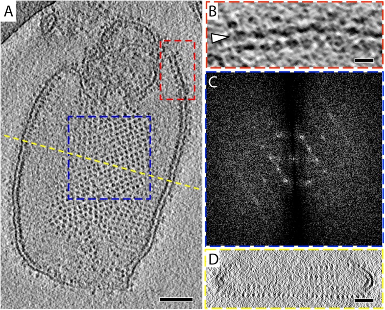
Figure 1. 2D crystals of F1Fo ATP synthase in vitreous ice.

(A) Tomographic slice of a vesicle reconstituted with F1Fo ATP synthase. The ATP synthases appear as 10 nm spherical densities located 15 nm above the membrane. (B) Enlarged view of red boxed area in A showing the zigzag membrane structure (arrowhead). (See also Figure 2.) (C) Fourier transform of the blue-boxed area in (A). (D) Cross-section along the yellow dashed line in (A). Scale bar: (A) 100 nm, (B) 20 nm, (D) 50 nm.

DOI: http://dx.doi.org/10.7554/eLife.06119.003

Figure 1.

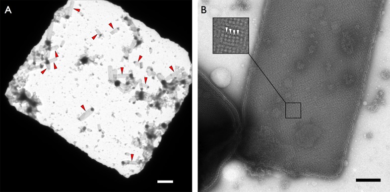
Figure 1—figure supplement 1. Crystalline vesicles of F1Fo ATP synthase in negative stain.

Figure 1—figure supplement 1.

(A) Overview of an EM grid square with numerous rectangular crystalline vesicles (red arrowheads). Scale bar 3 μm. (B) Rectangular crystalline vesicle at higher magnification with boxed area enlarged. F1 heads (white arrowheads). Scale bar 200 nm.
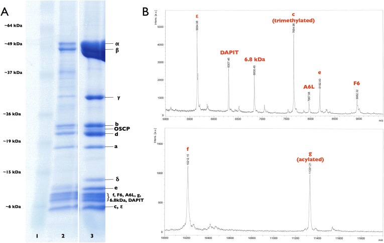
Figure 1—figure supplement 2. Subunit composition of F1Fo ATP synthase isolated from 2D crystals.

Figure 1—figure supplement 2.

(A) SDS-polyacrylamide gradient gel (10–20%) showing the subunit composition of purified F1Fo ATP synthases before (lane 2) and after (lane 3) 2D crystallisation. Lane 1, molecular marker (BenchMark). (B) Mass spectrometry profile of small subunits (5000–12,000 Da) of the F1Fo ATP synthase isolated from 2D crystals. All F1Fo ATP synthase subunits are present in the 2D crystals.
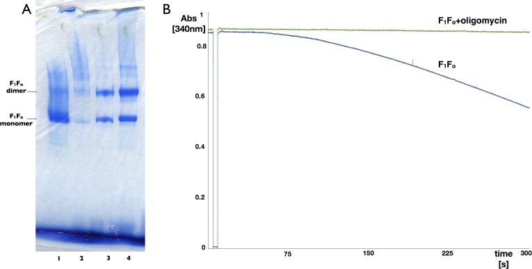
Figure 1—figure supplement 3. Blue-native polyacrylamide gel and activity assay of F1Fo ATP synthase isolated from 2D crystals.

Figure 1—figure supplement 3.

(A) BN polyacrylamide gradient gel (4–12%) of purified F1Fo ATP synthase solubilized in decylmaltoside (lane 1 and 2, 15 μg and 2 μg respectively) and of digitonin-resolubilised 2D crystals (lane 3 and 4, 30 μg and 90 μg respectively). Bands of lower molecular weight subcomplexes, that is, Fo or c8, are weak or absent. The dimer band in lane 1 and 2 presumably stems from mitochondrial F1Fo ATP synthase dimers preserved by mild purification conditions. The dimer band visible in lane 3 and 4 most likely represents non-physiological F1Fo ATP synthase dimers that form in the 2D crystals. (B) Typical ATPase activity and oligomycin-sensitivity of digitonin-resolubilised F1Fo ATP synthase isolated from 2D crystals. The hydrolysis of ATP by the F1Fo ATP synthase was monitored using an enzyme couple assay by detecting NADH oxidation at 340 nm at 20°C in the absence (blue) or presence (green) of oligomycin. Oligomycin inhibits ATP hydrolysis through F1 by stopping rotation of the Fo motor, that is, oligomycin sensitivity gives a measure of the amount of coupled, intact F1Fo ATP synthase complexes in the preparation, in this case >95%.