Figure 1.

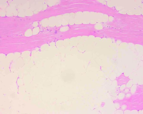
Intramuscular lipoma specimen. Various degrees of muscle atrophy, as well as pyknotic nuclei or nuclear swelling, shown in the specimen (hematoxylin-eosin stain, ×200).

Intramuscular lipoma specimen. Various degrees of muscle atrophy, as well as pyknotic nuclei or nuclear swelling, shown in the specimen (hematoxylin-eosin stain, ×200).