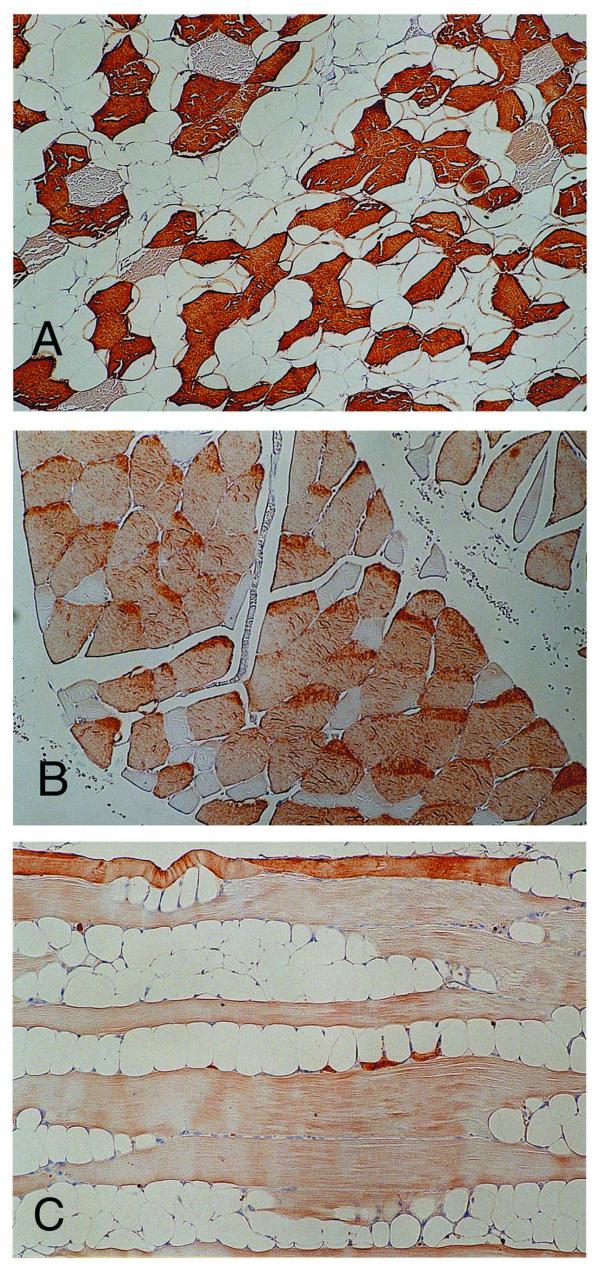
Figure 2.

Immunohistochemical study for the involving muscle fibers. (A) SERCA1-positive fast muscle tissues are dominantly preserved, with the other type prominently decreased (×125). (B) Slow muscle fiber tissues reacting to SERCA2 are conserved, and SERCA2-negative fibers are smaller and angulated (×125). (C) Fast muscle tissues stained by troponin T are shown dominantly atrophic and replaced with fatty tissues (×125).
