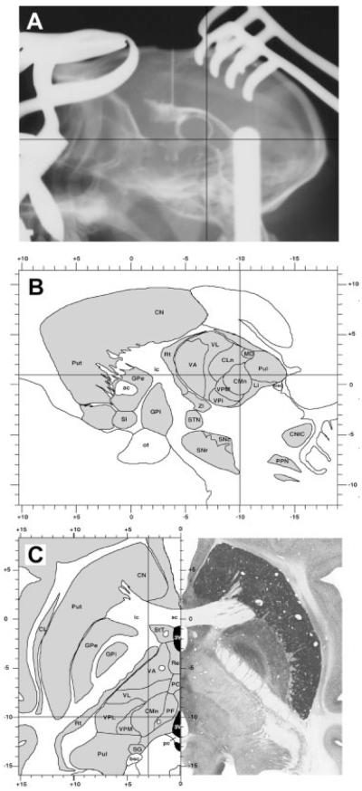
FIG. 1.
Ventriculography-assisted stereotaxic surgery targeting the centromedian thalamic intralaminar nucleus (CM). (A) Sagittal projection from an Rx plate showing the complete filling of the ventricular system as well as the coordinates for approaching the CM nucleus. (B, C) Stereotaxic coordinates for the CM nucleus, calculated according to a stereotaxic atlas of our own. Selected coordinates are illustrated in sagittal (B) and horizontal (C) brain maps of the primate Macaca fascicularis.

