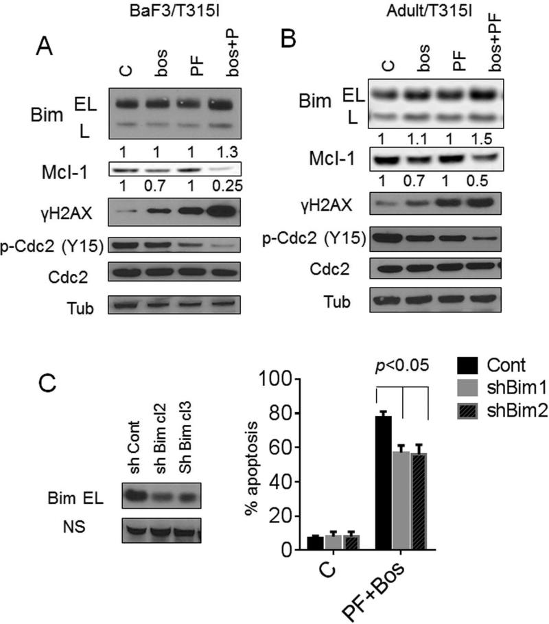
Figure 3. Bosutinib/PF induces Bim up-regulation and Mcl-1 down-regulation while promoting p34cdc2 activation and DNA damage.
BaF3/T315I (A) and Adult/T315I (B) cells were treated with 1.4 μM bosutinib alone or with PF (0.4 μM) for 36 hr after that cells were lysed and applied to Western blot. Expression of the indicated proteins was determined by Western blotting using the indicated antibodies. Each lane was loaded with 25 μg of protein; blots were stripped and reprobed with antibodies to tubulin to ensure equivalent loading and transfer. Values below the blots indicate densitometric determinations relative to untreated controls. (C) BaF3/T315I cells were stably transfected with shRNA-Bim or shRNA-control constructs in a pSUPER.retro vector. Two clones displaying reduced endogenous expression of Bim were isolated and used in subsequent experiments. The cells were exposed to PF/bosutinib for 72 hr, after which the percentage of apoptotic cells was determined by 7-AAD+ (p < 0.05, significantly greater than values for single-agent treatment). All values represent the means ± SD for duplicate determinations performed on 3 separate occasions.

