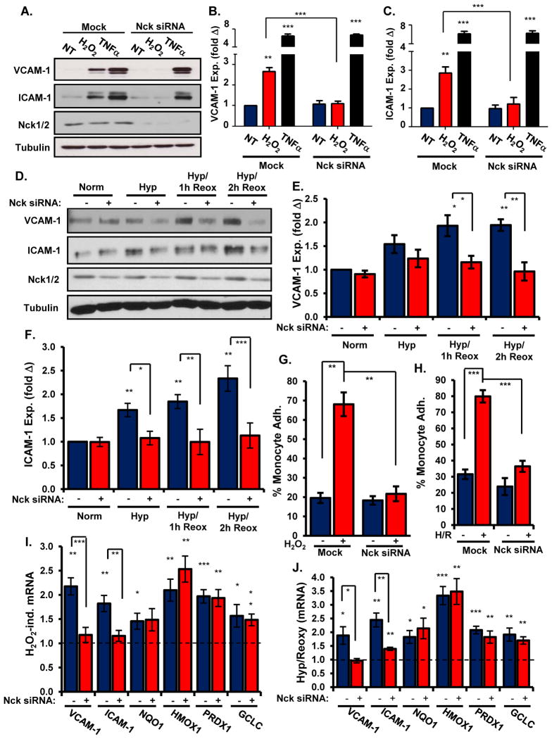
Figure 2. Depleting Nck expression blunts oxidant stress-induced endothelial activation and leukocyte recruitment.
(A–C) The abundance of VCAM-1 (A, B) or ICAM-1 (A, C) in HAECs transfected with Nck1/2 siRNA and treated with H2O2 or TNFα was determined by Western blotting. (D–F) The abundance of VCAM-1 (D, E) or ICAM-1 (D, F) in HAECs transfected with Nck siRNA and exposed to hypoxia/reoxygenation was determined by Western blotting. (G/H) HAECs transfected with Nck1/2 siRNA were exposed to H2O2 (G) or hypoxia/reoxygenation injury (H). The adhesion of Cell Tracker Green-labeled THP-1 monocytes was determined in static adhesion assays (fig. S3) and conveyed as percent of monocytes adhering under each condition. (I/J) HAECs transfected with Nck1/2 siRNA were exposed to H2O2 (I) or hypoxia/reoxygenation (J). Changes in the expression of genes encoding proinflammatory and antioxidant factors were determined by qRT-PCR. Results show the fold change in gene expression compared to untreated conditions. n=5 independent experiments in all panels. * p<0.05, ** p<0.01, *** p<0.001.

