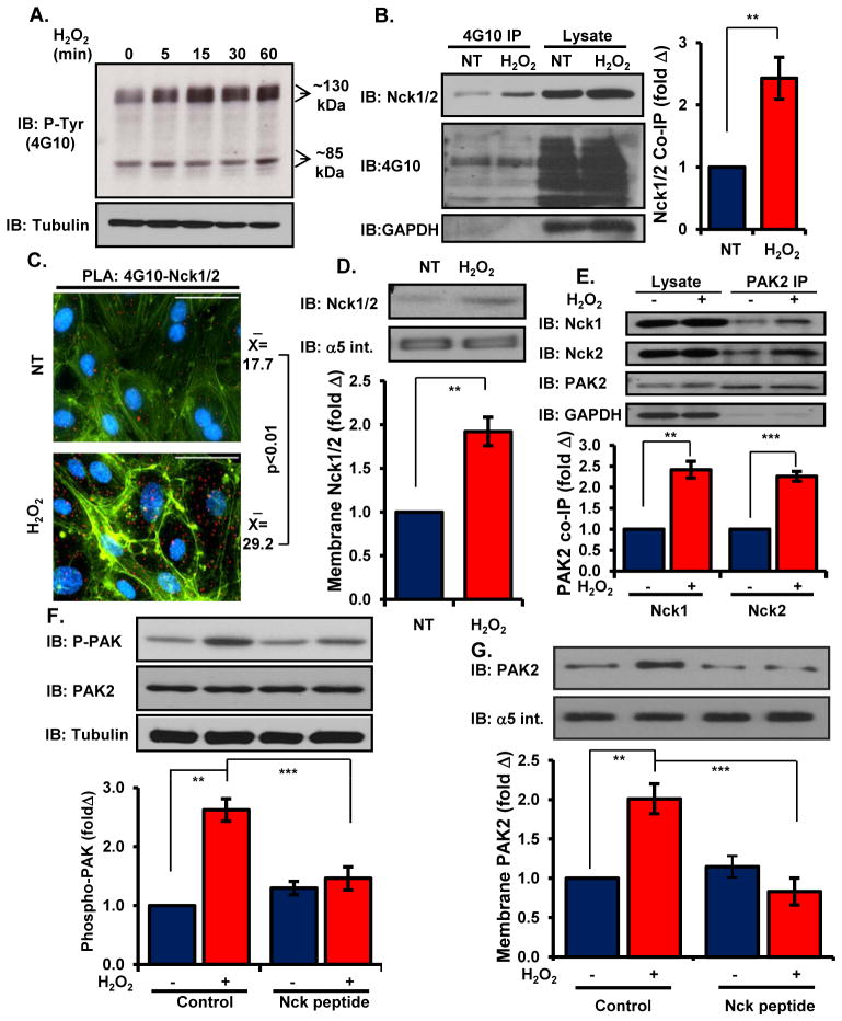
Figure 3. Nck couples oxidant stress-induced tyrosine phosphorylation to activation of PAK2.
(A) Total cellular tyrosine phosphorylation was determined in cells treated with H2O2 for the indicated times. Representative blots are shown. (B) Nck co-immunoprecipitation with tyrosine phosphorylated proteins was determined in cells treated with H2O2. (C) HAECs were treated as in (B) and Nck/phosphotyrosine interactions were analyzed in situ by proximity ligation assays. Average proximal ligations per cell are shown. (D) Cells were treated as in (B), and Nck recruitment to the membrane fraction was determined by Western blotting and normalized to the integral membrane protein α5 integrin. (E) HAECs were treated as in (B) and PAK2 co-immunoprecipitation with Nck1 and Nck2 was analyzed. (F) Activation of PAK2 was determined by Western blotting in cells pretreated with Nck-blocking peptide before treatment with H2O2. (G) Cells were treated as in (F), and PAK2 recruitment to the membrane fraction was determined by Western blotting and normalization to the integral membrane protein α5 integrin. n=5 independent experiments for each panel. * p<0.05, ** p<0.01, *** p<0.001.

