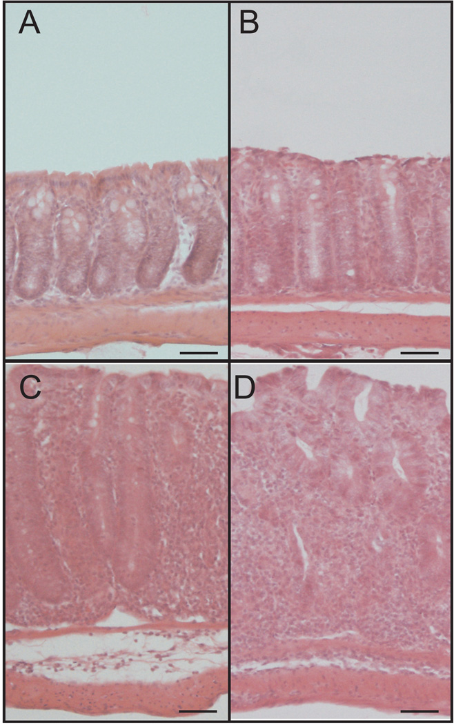
Figure 1. T cell transfer colitis-mediated tissue histopathology over time.
A–D: T cell transfer-mediated histological changes illustrated by hematoxylin and eosin staining of tissue harvested at weeks 0, 2, 4 and 6, respectively. Histopathological changes progressively increase over time including neutrophil infiltrate, abnormal crypt architecture, goblet cell loss, and degree of inflammation infiltrate in the lamina propria. Magnification is 200×. Scale bars, 50µm.

