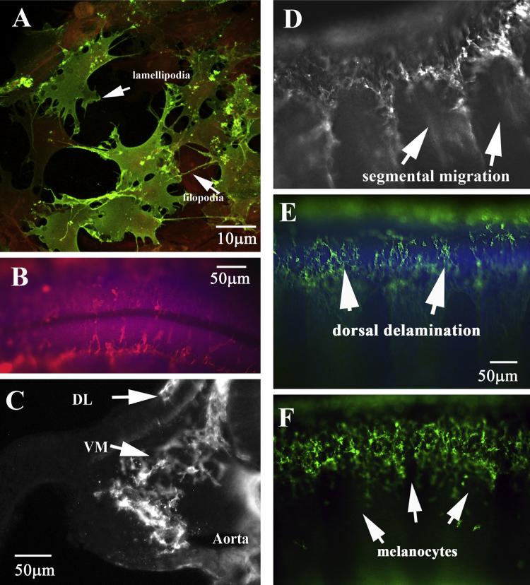
Fig. 1.
HNK1 labels chicken neural crest cells in great detail. (A) Neural crest cells in culture show classic morphology of mesenchymal cells: many filopodia and lamellipodia. Neural crest cells are stained with HNK1 (green) and other cells with actin (red). (B) Dorsal view of delaminating trunk neural crest cells on top of the two halves of the trunk neural tube. The neural tube is labeled with DAPI. (C) Midtrunk section through HH17 embryo shows migrating trunk crest cells throughout the trunk region. (D–F) Wholemount side views of migrating trunk neural crest cells in caudal (D) or rostral (E) of HH17 embryo and rostral (F) of HH20 embryo. DL: dorso-lateral migrating NCC, VM: ventro-medial migrating NCC. (For interpretation of the references to color in this figure legend, the reader is referred to the web version of this article.)

