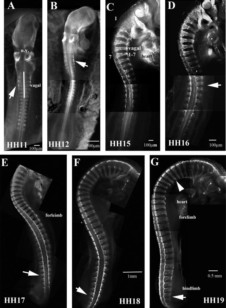
Fig. 2.
Wholemount views of HH11–19 embryos with HNK1. HNK1 labels migrating trunk neural crest poorly in HH11 and HH12 (A, B), but strongly by HH15 (C). Footnotes indicate the stage of the chicken embryo. Vagal crest is indicated in (C). Arrow in (D) points to 16th somite. Arrow in (E–G) to migrating NCC at tail ends. Arrowhead in (G) point to thinning of NCC streams, indicating that they are beginning to condense. In (A) o.v. stands for otic vesicles.

