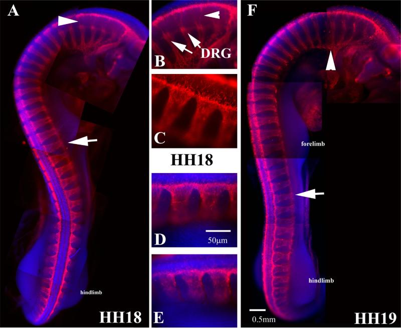
Fig. 6.
Wholemount views of HH18 and HH19 embryo with HNK1. (A) Wholemount embryo with HNK1 shows the peak of trunk neural crest migration along the whole trunk in HH18 embryo. (B–E) Higher magnification of the rostro to caudal progression of neural crest migration of HH18 embryo in (A). DRGs can be seen in (B) (arrows). (F) HH19 embryo, arrowhead points to vagal region spinal nerves, arrow points midtrunk crest cells still migrating.

