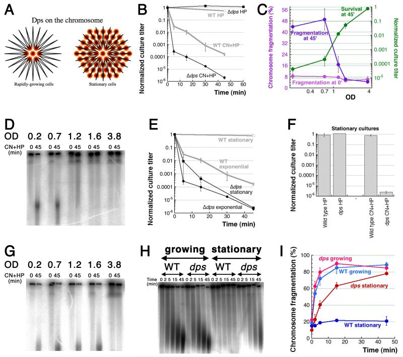
Fig. 5. Resistance of stationary cells to CN+HP is due to Dps.
A. Dodecameric Dps protein forms spheres that bind DNA non-specifically and accumulate up to 500 iron atoms per sphere. Orange/brown small circles, Dps dodecamers; black line in the shape of a star, chromosomal (duplex) DNA. B. Kinetics of killing by the two treatments of Δdps mutant. WT curves from Fig. 1C are shown in grey for comparison. C. OD-dependence of CN+HP killing and fragmentation of wild type cells. Aliquots of the same culture were challenged with standard CN+HP treatment at various ODs of the culture, and their normalized survival or fragmentation were plotted as a function of OD. In our growth conditions, the maximal density (fully stationary cultures) of WT cells does not exceed 4.0. D. A representative pulsed-field gel of OD-dependence of fragmentation in WT cells after CN+HP treatment. E. Kinetics of CN+HP killing of exponential vs stationary cultures of wild type cells and Δdps mutant. F. Stationary cultures killing by HP-alone or CN+HP 45 minute treatment. G. OD-dependence of fragmentation in Δdps mutants: a representative gel, similar to the one in (D). Cultures were treated without prior dilution. H. A representative pulsed-field gel of CN+HP treatment kinetics to show that Δdps stationary cultures undergo robust chromosomal fragmentation if diluted 10× into a fresh medium right before the treatment. I. Kinetics of chromosome fragmentation in wild type and Δdps cells, either in growing or stationary/diluted cultures. This is quantification of several independent runs like in “H”.

