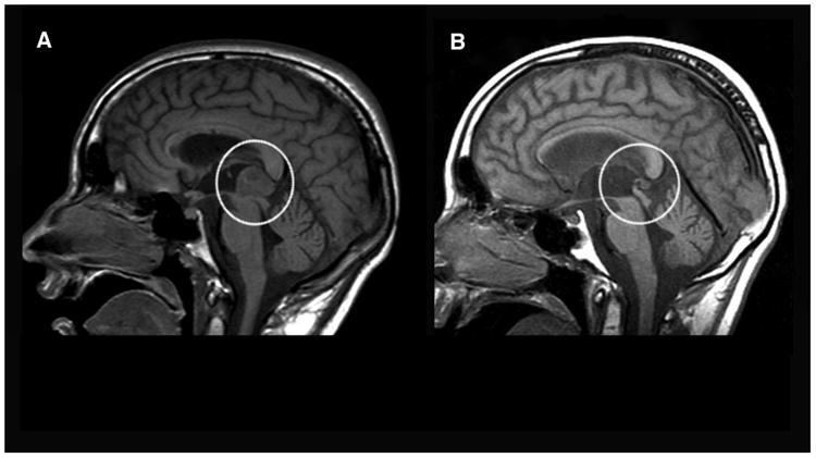
Figure 1.

Baseline and 16-Month Follow-up Magnetic Resonance Images Demonstrating Pineal Region Mass Before and After Chemotherapy Treatments and Stem Cell Transplanta.
aThe scan image presented in “A” represents the soft tissue mass in the dorsal midbrain/pineal cistern (circled; 29mm). The image represented in “B” shows the post-treatment rescinded pineal germ cell tumor 16 months later (circled; 3.7mm).
