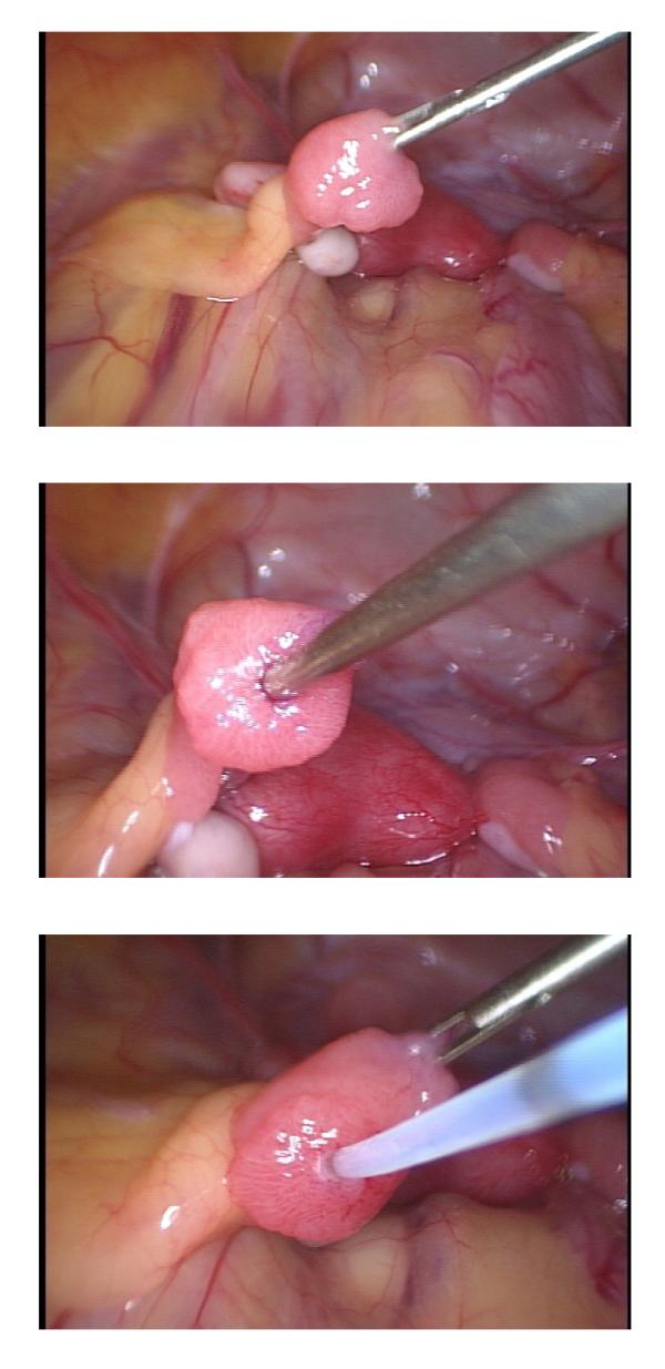
Figure 3.
Illustrations of the progressive stages in laparoscopic embryo transfer in rhesus monkeys. In Panel A, the fimbrium is grasped with a Patton retractor and placed in traction. A guide cannula is then introduced into the oviduct (Panel B). Finally the loaded transfer catheter is inserted transabdominally and advanced into the oviduct 1–3 cm in preparation for embryo deposition (Panel C).

