
Fig. 2.
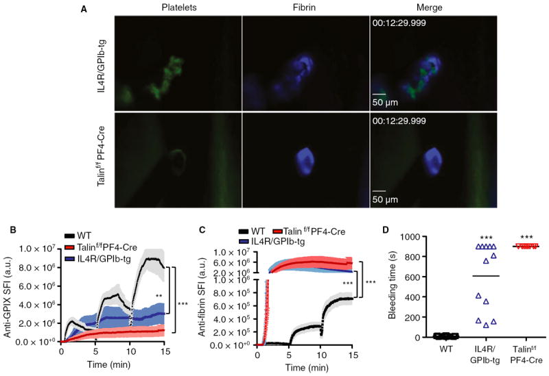
Contribution of platelets to hemostasis after laser injury. Vascular lesions were induced in wild-type (WT) (black line/symbols), talin1f/fPF4-Cre (red) and IL4R/GPIb-tg (blue) mice. (A) Representative images. (B, C) Accumulation of platelets (B) and fibrin (C). Sum fluorescence intensity (SFI) ± standard error of the mean is shown. Talin1f/fPF4-Cre: n = 9 (four mice). IL4R/GPIb-tg: n = 10 (four mice). (D) Bleeding time. **P < 0.001, ***P < 0.0001.