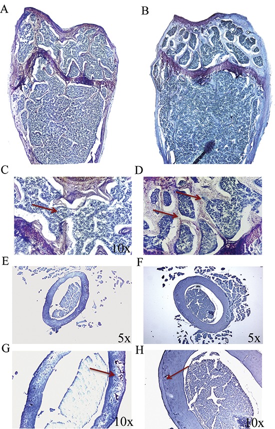
Figure 4. p62 DNA rescues osteoporosis.

Representative reconstructions of metaphyseal regions of distal femurs from OVX-pcDNA3.1 (A, C) and OVX-p62DNA mice (B, D). Arrows indicated the trabecular bone loss (C) and the restored trabecular microarchitecture (D) Representative sections of femur mid diaphysis from OVX-pcDNA3.1 (E, G) and OVX-p62 DNA mice (F, H). Note the expansion of the medullary cavity and the resorption cavities within the cortex (Arrow, G). Arrows indicated the reconstitute cortical bone structure (H) Magnifications: 10 × (C, D, G, H), 5 × (E, F).
