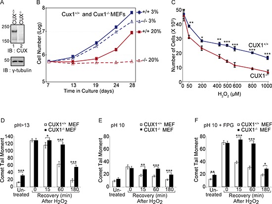
Figure 1. Genetic inactivation of Cux1 causes a proliferation block in atmospheric (20%) oxygen.

(A) Immunoblotting analysis using CUX1–1300 antibody. (B) Cux1+/+ and Cux1−/− MEFs were cultured in 3% or 20% oxygen and counted over a period of 21 days. (C) Cux1+/+ and Cux1−/− MEFs were maintained in 3% oxygen for 7 days and exposed to various concentration of H2O2 for 60 min and then cultured in fresh medium for 24 h in 3% oxygen before counting the cells. Error bars represent standard error. *p < 0.05, **p < 0.01, ***p < 0.001; Student's t-test. (D, E and F) Cux1+/+ and Cux1−/− MEFs were maintained in 3% oxygen for 7 days, exposed to 50 μM H2O2 for 20 minutes and allowed to recover for the indicated time. Cells were submitted to single cell gel electrophoresis at pH > 13 (D), pH 10 (E), and pH 10 in the presence of FPG (F) Comet tail moments were scored for at least 50 cells per condition. Error bars represent standard error. *p < 0.05, **p < 0.01, ***p < 0.001; Student's t-test.
