
Figure 1. Brief treatment with H2O2 induces premature senescence in HEI-OC1 cells.
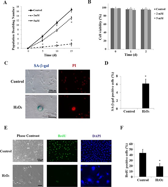
(A) Population doubling time. Population doubling experiments were performed in duplicate, as described in the Methods. (B) Cell viability was determined by trypan blue staining at the indicated times after brief (1 h) treatment with H2O2 (2 mM and 5 mM). All values are means ±S.D. from three or more independent studies. *P < 0.05. (C) Representative senescence-associated β-galactosidase (SA-β-gal) staining of HEI-OC1 cells. Propidium iodide (PI) staining labeled nuclear DNA. The assay was carried out in duplicate 2 days after completing the treatment described in the Methods (Cell Viability Assay section). (D) SA-β-gal-positive cells were quantified by counting more than 100 cells for each sample. All values are means ±S.D. from three or more independent studies. The control condition exhibited no detectable SA-β-gal staining; *P < 0.05. (E) Representative bromodeoxyuridine (BrdU) assay results conducted 2 days after brief treatment with H2O2 (5 mM for 1 h). DAPI was used to counterstain DNA in the nucleus for cell identification. (F) BrdU positive HEI-OC1 cells were quantified by counting more than 100 cells for each sample. Values are means ±S.D. from three or more independent studies. *P < 0.05.