
Figure 2. Effects of brief H2O2 treatment on autophagy signaling pathway in HEI-OC1 cells.
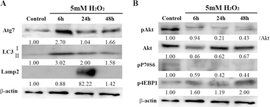
(A) Representative Western blots showing the expression of autophagy-related 7 (Atg7), macrotubule-associated protein 1 light chain 3 (LC3), and lysosome-associated membrane protein 2 (Lamp2) in control and H2O2-treated cells. β-actin was included as a loading control. The optical density of Atg7, LC3 II, and Lamp2 in H2O2-treated cells was normalized to that of the corresponding proteins in control cells, and the resulting ratios are indicated below each blot. (B) Western blot analysis of the mammalian target of rapamycin (mTOR) pathway, which negatively regulates autophagy. This pathway includes phosphorylated Akt (pAkt), Akt, pP70S6, and p4EBP1. β-actin was included as a loading control. The optical density of pAkt, Akt, pP70S6, and p4EBP1 in H2O2-treated cells were normalized to that of the corresponding proteins in control cells, and the resulting ratios are indicated below each blot.