
Figure 5. AMPK dysfunction resulting from AMPKα knockdown leads to premature senescence in HEI-OC1 cells.
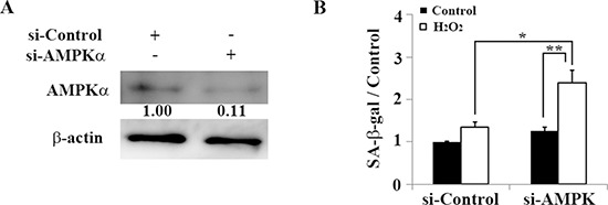
(A) Representative Western blot showing AMPKα expression in HEI-OC1 cells transfected with control or AMPKα siRNA. AMPKα expression levels were determined 2 days after transfection to validate the efficiency of AMPKα knockdown. β-actin was included as a loading control. The optical density of AMPKα in H2O2-treated cells was normalized to that of AMPKα in si-control cells, and the resulting ratios are indicated below each blot. (B) Quantification of SA-β-gal staining intensity 2 days after 1-h treatment with 5 mM H2O2 and transfection with control siRNA or AMPKα siRNA. Quantitative fluorescence analysis was conducted as described in the Methods. All values are means ±S.D. from three or more independent studies. *P < 0.05; **P < 0.01.